The Orthopedic TMJ Blueprint: Airway Focused Reconstructive Surgery
Why jaw reconstructive surgery is fundamentally airway surgery, the limits of dental sleep appliances, and how orthopedic anchor stitches protect live joints.
Shifting from Soft Tissue Myths to Hard Bone Realities
For a long time, the medical community believed that sleep apnea was just a soft tissue issue. Doctors assumed that a floppy palate or an oversized tongue was blocking the throat. But modern airway science has proven something completely different: the underlying bone structure dictates exactly where soft tissue blockages happen.
Think of it like a tent. If the wooden poles are too short or collapsed, the canvas fabric collapses into a messy heap. In the human throat, if the maxilla or mandible is underdeveloped, the structural poles are too small. This structural deficiency pulls all the attached throat muscles out of alignment, directly choking off the airway space.
True jaw reconstructive surgery is fundamentally airway surgery. Moving the physical bones forward does not just fix a patient's bite or alter their appearance. Moving the skeletal structure stretches the surrounding deep muscles, creates ideal tissue tension, and instantly expands the patient's breathing paths.
The Three Hidden Throat Spaces and Advanced Orthopedic Stitching
In this clinical session, world-renowned oral and maxillofacial surgeon Dr. Scott Boulding joins host Dr. Melissa Seibert to share the essential diagnostic rules for screening airway obstructions and fixing damaged jaw ligaments.
The Three Airway Spaces Every Dentist Must Check
When screening a patient for breathing or sleep disorders, you cannot treat the throat as one big tube. You must separate the airway into three distinct geographic levels using a standard 3D scan:
- The Nasopharyngeal Space: This sits at the very back part of the nose. If a patient has a narrow, underdeveloped upper jaw, this nasal zone gets heavily constricted. Expanding this space using bone advancement surgery is one of our most predictable ways to cure sleep apnea entirely.
- The Oropharyngeal Space: This lives directly behind the tongue. Its size is determined by the horizontal position of the lower jaw. Moving the lower jaw forward instantly pulls the tongue base away from the throat wall.
- The Laryngopharyngeal Space: This is the deepest zone down by the epiglottis and windpipe. If the lower jaw lacks proper length, the hyoid bone drops down and slips backward, completely crushing this deep breathing track.
The Unintended Side Effects of Dental Sleep Appliances
When a patient cannot tolerate a CPAP machine, general dentists frequently prescribe a removable mandibular advancement appliance. While these dental mouthpieces are widely popular, they come with significant structural risks. A sleep appliance only opens up one zone: the middle oropharyngeal space behind the tongue. If the patient's true blockage lives up in their nose or deep down by their throat base, a dental appliance fails to help them breathe.
Worse, wearing these devices long-term places intense, unnatural stress on the temporomandibular joint. Forcing the lower jaw forward stretches the delicate joint discs and ligaments all night long. Dr. Boulding regularly treats patients who have developed severe jaw pain, clicked discs, and permanent bite shifts where their back teeth no longer touch because of sleep appliances.
Fixing Torn Ligaments with Orthopedic Anchors
When a joint disc slips permanently out of position, it means the surrounding structural ligaments are torn or stretched. Without an active cushion, the joint loses its vital pool of lubricating synovial fluid. This lubrication loss causes the raw bone to grind against bone, leading to rapid condyle erosion and bone spurs.
To repair this damage, modern surgeons rely on precise orthopedic principles. Using a tiny, minimally invasive access incision, the slipped disc is carefully pulled back onto the head of the condyle. Then, the surgeon reinforces the torn posterior and lateral ligaments using a permanent orthopedic anchor stitch. This specialized thread completely incorporates into the living tissue, making the ligament naturally stronger so it can hold the disc in place forever.
Clinical Takeaways
- Focus on Bone Foundation: Realize that underlying skeletal position directly dictates soft tissue collapse and airway constriction in adults.
- Map Three Separate Spaces: Use a standard 3D office scan to evaluate the nasopharyngeal, oropharyngeal, and laryngopharyngeal zones individually.
- Monitor Sleep Appliance Side Effects: Screen mandibular advancement patients for long-term joint pain, stretched ligaments, and lost posterior tooth contact.
- Order Joint MRIs Early: Skip standard x-rays for severe joint pain; rely on an MRI as the gold standard to diagnose hidden ligament tears.
- Stitch the Support Ligaments: Reinforce displaced joint discs by securing them with permanent orthopedic anchors that fuse directly into the living tissue.
Chapters & Timestamps
| Timestamp | Topic Covered in Episode |
|---|---|
| [00:00] | Introduction and Accessing the Free Step-by-Step Injection Molding PDF |
| [04:30] | Airway Reconstructive Surgery: Maximizing Three Throat Spaces |
| [15:10] | Why Moving Bones Succeeds Where Soft Tissue Cutting Fails |
| [26:45] | The Appliance Trap: How Mandibular Devices Damage Jaw Joints |
| [38:20] | Standardizing Office Scans: Using CBCT as a Patient Screening Tool |
| [46:15] | Why MRIs are the Ultimate Medical Gold Standard for Ligament Tears |
| [54:30] | Orthopedic Anchors: Stitching Discs and Handling Post-Surgical Bites |
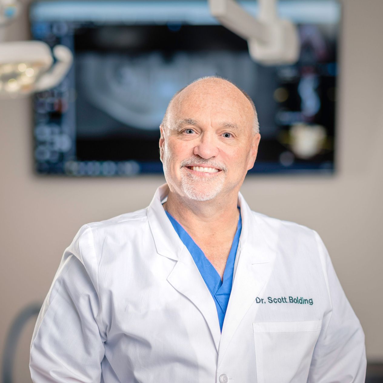
Dr. Scott Boulding
Oral & Maxillofacial Surgeon
An internationally renowned oral and maxillofacial surgeon practicing in Florida and Arkansas who is recognized internationally for his integration of joint, facial, and airway optimization. Dr. Boulding has dedicated his career to airway-focused orthognathic reconstruction and advanced TMJ surgery. He lectures worldwide on advanced data models, teaching contemporary surgical workflows that treat the full human behind the smile.
Surgical Practice Systems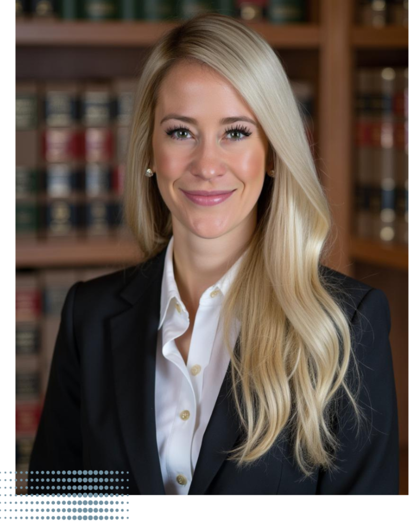
Dr. Melissa Seibert
DMD, MS, FAGD, ABGD
Creator and host of the Dental Digest Podcast — the #1 clinical dental podcast worldwide and a top 1% global podcast. Dr. Seibert is a former active-duty U.S. Air Force dentist, internationally sought-after speaker, Key Opinion Leader, and published author in top dental journals. She is passionate about equipping general dentists with high-level, evidence-based clinical skills.
Publications & SpeakingMaster This Workflow in Your Practice
The Dental Digest Podcast brings you the theory — but Elevated GP gives you the over-the-shoulder execution. Step-by-step video masterclasses, clinical mentorship, and CE credit to implement these techniques seamlessly. Join our global community of dentists.
Explore the Elevated GP MembershipStudies & Resources
- Boyd, S. B., Walters, A. S., Waite, P., Harding, S. M., & Song, Y. (2015). Long-Term Effectiveness and Safety of Maxillomandibular Advancement for Treatment of Obstructive Sleep Apnea. Journal of Clinical Sleep Medicine, 11(07), 699-708.
- Zhou, N., Ho, J. P. T. F., Spijker, R., Aarab, G., de Vries, N., Ravesloot, M. J. L., & de Lange, J. (2022). Maxillomandibular Advancement and Upper Airway Stimulation for Treatment of Obstructive Sleep Apnea: A Systematic Review. Journal of Clinical Medicine, 11(22), 6782.
- Walker, A., Kassir, M. F., Sama, V., Nguyen, S. A., & Abdelwahab, M. (2025). Maxillomandibular Advancement Safety and Effectiveness in Obstructive Sleep Apnea: Systematic Review and Meta-Analysis. Otolaryngology–Head and Neck Surgery, 172(4).
- National Institutes of Health (NIH). (1996). consensus Development Conference Statement on Management of Temporomandibular Disorders. Journal of the American Dental Association, 127(11), 1595-1603.
- Free Clinical Resource — Step-by-Step Anterior Injection Molding Technique Guide
Full Episode Transcript
Dr. Melissa Seibert: Hey, I want to tell you about something I put together for you. I created a free PDF guide that walks you step-by-step through the injection molding technique. I love this technique because it is one of the best ways to get predictable, beautiful, and highly aesthetic anterior composites. You're actually injecting the composite directly into the tooth using a clear template which makes it far more consistent and efficient. If you like this guide, I've made it super easy. Just head over to theelevatedgp.com forward slash IMPDF. And to make it even simpler, I've included the link for you right here in the show notes.
Hey, welcome to Dental Digest. I'm your host, Dr. Melissa Seibert. This is a podcast with a mission of enabling you to stay on the cutting edge of evidence-based dentistry. This is part one of a two-part series, and you're going to want to be sure to come back next week for part two. By the way, if you hear a bit of an impediment, it's because I'm going through comprehensive ortho. Joining me today is Dr. Scott Boulding, an oral maxillofacial surgeon whose career has been dedicated to TMJ surgery and airway-focused orthognathic reconstruction. He practices in both Florida and Arkansas, lectures internationally, and is widely recognized for his integration of joint, facial, and airway optimization into contemporary surgical workflows. In this episode, we explore Dr. Boulding's philosophy that orthognathic surgery is fundamentally airway surgery, why skeletal positioning dictates soft tissue obstruction, and how mandibular advancement can dramatically transform patient physiology. We also discuss how he evaluates the nasopharyngeal, oropharyngeal, and laryngopharyngeal spaces using CBCT and DICE, and how he determines where true obstruction originates.
I've got something that you are going to love. If you've been loving the podcast and want to dive deeper into mastering implant dentistry, this is your chance. I'm giving away access to one of my on-demand courses completely free. This course is packed with everything you need to know about implant occlusion, platform switching, and creating that stunning aesthetic emergence profile that sets your work apart. Here's how to grab it. Leave a rating for the podcast, take a quick screenshot, and send it my way. You can either direct message me on Instagram, my Instagram handle is dr.melissaseibert, or send it to me in an email at dr.melissaseibert at gmail.com. Once I get the screenshot, I will send you access to the course so you can start learning right away. It's my way of saying thank you for supporting the podcast and being part of this amazing community, and I can't wait to hear what you think.
So let me ask you this, when you are structuring orthognathic cases, some orthognathic surgeons might be very aesthetically forward, where they're effectively planning the course of surgery based on achieving the most aesthetic outcome. Others might be more joint-centric, and then others might be more airway-centric, and then a combination thereof. Where is your stance on this?
Dr. Scott Boulding: Melissa, it's on all the above. So if you look at orthognathic surgery, orthognathic surgery is jaw reconstructive surgery, right? And the reason we're doing that surgery is to position generally the maxilla, the mid-face, the nose, the mandible, the tongue, the palate, the chin, the hyoid bone in the proper position. And what I've found through the years now, especially in the last five or six years, that we've really been focusing on airway health and improving airway and helping patients get off CPAP machines. We've noticed that, okay, the main reason we need to do this surgery is to really get a good, well-balanced, and rounded opening, both in the nasopharyngeal area, the oropharyngeal area, and the laryngopharyngeal area. So there's three spaces in the back of the throat that we want to grow and make sure it's larger. And when you have obstructive sleep apnea or you have air issues, it can be any of those spaces that can be the obstruction, or it can be all three, depending on what the defect is. But we've got to remember that obstructive sleep apnea is primarily and predominantly associated with an obstruction, and that obstruction used to be thought with soft tissue, but it's the bone that determines where the soft tissue obstruction occurs.
So back to your question, if we do orthognathic surgery and we're not focusing on the airway, and now that we know that the airway is very important to overall health as a population, then we're not doing the patients a proper service. Because if we get the face what we think is aesthetically right, but we miss the airway, then we've not really helped the patient overall. As a matter of fact, we could have made it worse. So it's important to make sure that the airway is the primary focus. Secondarily now that we know aesthetics is important to all of us, if we properly position the maxilla in relation to the face and angle the posterior maxilla in the proper direction, whether we do a counterclockwise rotation or a clockwise rotation, and we make sure that we get the tooth to lip properly oriented with the smile and the anterior positioning and the chin position, the aesthetics goes along with it. And so what I'm finding, and that's what I teach my residents and my fellows, if we get the airway right and balanced properly, the face is going to look right.
And so, you know, when we first started doing these for airways, we were worried that we were going to go through the cephalometric norms of aesthetics. But what I found is when we tried to treat to the cephalometric norms that I was taught early in my career, I wasn't getting the airway where it needed to be. So we started blowing through those traditional valued lines to open the airway. And so I was worried. I was worried I was going to make everyone look like a chimpanzee. But in reality, what happened is the patients look much healthier. And then we're talking patients that we move the maxilla for 10, 12, 13 millimeters and still have really good aesthetics. So to answer your question, I really look at joints, look at airway and look at faces. It's very important. I think as a surgeon, we need to do all three. And I don't think you can focus just on one.
Dr. Seibert: You made a really interesting statement. And you said that historically, we thought that the obstruction was coming from soft tissue. But you said that the bone determines the obstruction. Tell us more about that.
Dr. Boulding: So when you look in the back of the throat for patients, you look at the lateral pharyngeal wall, the posterior pharyngeal wall, look at the tongue base, and you're looking at essentially the airway for patients. In the early days, the treatment was not considered to do jaw surgery. If you had obstructive sleep apnea, of course, the number one treatment is CPAP today. In the early days, the otolaryngologist would try to open the soft tissues by doing soft tissue surgeries, either doing tongue reduction surgery or tongue advancement surgery or UPPP where they take off the palate and open up the airway. And while these would get some early good results, these tissues happened to regrow and relapse. So the success of these soft tissue surgeries was not as successful. As a matter of fact, they were abandoned by many ENTs for treating these patients. Plus, it was a very difficult surgery, very bloody surgery, very painful surgery.
Now that we know that we have the stability that we have with custom plates and BSP planning with jaw surgery, by moving the bones, you're actually stretching those tissues that open the airway. And those tissues that we were trying to reduce, if you move the bones, they actually go into the proper position and you get better tension on all these muscles, which opens up the orifice, opens up the airway. And so when you look at the DICE studies, both before and after reconstructive surgery, you can see dramatic changes in the soft tissue openings after you do the jaw surgery.
Dr. Seibert: You also talked about how there's multiple areas at which a person can obstruct. How do you identify which area or areas at which they're obstructing and how to plan your surgery accordingly?
Dr. Boulding: That's another very good question, Melissa. And so when we think about obstructive sleep apnea, when it's truly obstructive sleep apnea, not central sleep apnea, one should question in your mind, where is the obstruction? And the obstruction can be one of many places. Maybe it's just a normal jaw position and the patient has a severe deviated nasal septum. It could be just nasal that is actually obstructing or not allowing the patient to properly breathe. But it also can be the posterior aspect of the upper jaw and the nose. In the posterior aspect of the maxilla, we have the posterior choana, which is the back part of the nose. If you have an underdeveloped maxilla and a very narrow maxilla and it doesn't project forward enough, then the posterior aspect of the nasal complex is also narrow. So that's the nasopharyngeal space. The nasopharyngeal space is very important and is probably one of our most successful areas of treatment when we eliminate obstructive sleep apnea when doing orthognathic surgery or MMA. So the nasopharyngeal space airway is important. You can see that on CBCT. You can see that on a CT scan. You can also see that with DICE and with a simple nasal exam looking at the nasal septum.
Oropharyngeal is usually easier for us to see, especially on CBCT and CT. The oropharyngeal airway is behind the tongue and that's the tongue position based on where the position of the mandible is. We all know through CPR, moving the mandible forward brings the tongue forward and can open the oral airway. But the problem that we see, especially with sleep appliances, is many dentists think if the patient can't tolerate CPAP, the number two treatment recommended in the world is sleep appliances. The problem with sleep appliances is they only improve the oropharyngeal airway space. So when you bring the mandible forward, you may be helping the oropharyngeal airway space, but maybe they had a normal oropharyngeal airway space and you're not helping the patient at all. So I tell dentists that do recommend oral appliances, I think they are fine as long as it's treating the problem. So you need to both either titrate and test post-appliance just to make sure it's working.
The laryngopharyngeal space is down closer towards the hyoid bone and where the trachea and the epiglottis set. And that has to do a lot of times with the hyoid bone positioning. So if the hyoid bone is not suspended superiorly and anteriorly enough because the mandible's deficient, you can also have a collapse of the laryngopharyngeal space, which is down around the epiglottis. So you really need to evaluate, that's where DICE is very effective. So DICE is for your audience a laryngoscopy essentially with induced sleep via an IV sedation where we put a tube down the nose and kind of see where the obstruction is, and I look at all three spaces. What's the nasopharyngeal space look like? What's the oropharyngeal space? And if we can get down by the epiglottis, what does that space look like with inspiration? You can also see that on CBCT. And what I recommend for patients and for dentists if they have a CBCT in their office is just make sure the patient puts their tongue in the roof of their mouth right at the posterior aspect of their central incisors and try to take your CBCTs in that position in a standard position. And while it cannot be diagnostic, it can give you at least a screening tool to see if their airways are very narrow and you can look at all three of those spaces on the CBCT.
Dr. Seibert: How many of your cases are you using DICE for? Do you always do this as a screening tool? There's different convictions. Some will say that they think everyone should be doing it. Some say really only particular cases.
Dr. Boulding: I don't think it needs to be done on every case. If I have a patient that has obstructive sleep apnea and I have a CBCT and I can see where their spaces are, then I don't need that for a diagnosis. It's really important for patients that are undergoing treatment for, say, if they want the Inspire device, which is the little implantable device that helps the tongue move forward by attaching to the hypoglossal nerve. When you look through DICE, you can have different concentric constrictions of the pharyngeal airway. You can either have an anterior-posterior constriction or a lateral wall constriction or a full concentric constriction. If you have an anterior-posterior collapse, then the Inspire will work fine. If you have any of those other components, the Inspire will not work. If you use that modality for treating, absolutely you have to use the DICE procedure. The DICE procedure is a very good screening tool. I just don't think it's 100% necessary, at least when we're doing MMA work. It's a very low number of my patients I screen with it. I've considered doing it on every patient, but it's an extra cost. It requires the patient to go to sleep, and we've got to add that to another modality of treatment for the patient. But if it's a severe case and you want to know, I think it's a great tool.
Dr. Seibert: Yeah, you're almost creating more friction there for the patient. You mentioned mandibular advancement devices. Interested to hear your thoughts on this a little bit. My conviction, based on the preponderance of evidence, is that I really try to avoid them. I probably prescribe a mandibular advancement device once every two years or so. The preponderance of evidence just shows that this really can create occlusal changes in their bite. Some might ask, well, oftentimes they have these AM aligner devices. Couldn't this offset it? But that hasn't actually been extensively studied. I also think that there needs to be better patient education related to CPAPs. Patients might be unsuccessful the first few weeks, might experience some discomfort. I think there need to be better measures in place to perhaps train patients and equip them to get comfortable with it. I have been sleeping with a CPAP for seven years, and I will say it did take months to get comfortable with it. You're given a recipe, but their guidelines aren't typically very helpful. It's the same sort of guidelines that you can just get off of Google. I think we need better measures in place to also help patients potentially get comfortable with CPAP if surgery is not an option for them or a road that they want to go down.
Dr. Boulding: That a great thought, Melissa. Here's what I would tell you. We've got to remember, whether it's an anterior repositioning appliance, whether it's CPAP, or even whether it's Inspire, all three of those modalities of treatment for obstructive sleep apnea are not addressing the underlying problem in a potential cure fashion. So we've got to remember that MMA surgery, if you want to cure your disease potentially—and again we have to say potentially just to make sure we test post-surgically—but the only way you're ever going to really solve the problem is with surgery if you're an adult. As far as anterior repositioning appliances, to be honest with you, I'm not a big fan. Being a large TMJ surgeon, I see a lot of patients who have worn these different devices for many years or not even a long time and they develop TMJ symptoms. You've got to remember when you're repositioning the mandible forward, you're bringing the disc and the associated ligaments and everything and then you're stretching them nightly.
So while I think appliances work for pharyngeal airway problems, for example, I can't wear one. If I wear one, my jaw joints are completely sore the next morning and then my posterior teeth do not come together. And then if I bring my mandible forward enough to make my joints not sore, it's not effective in terms of my airway. So I'm not a big fan, but it's better than nothing, right? So you're talking to a guy that lost his father due to obstructive sleep apnea. So I'm very passionate about it. So if a patient can't tolerate CPAP and they just really can't do it and they're not willing to do surgery to correct it, then it's better than nothing. So I would say we just need to make sure that when we're doing those appliances, we understand what we're doing to the joints and understand what we're doing as far as bringing the oropharyngeal airway space, and make sure it's really working with post-appliance testing.
Dr. Seibert: Speaking of TM joint surgery, that is the whole reason I invited you on. You gave a presentation about ligament repair that I was so impressed by, a very contemporary approach to TM joint surgery. So the first thing we should talk about is, can you speak about this specific surgery where you have partial or complete ligament detachment and how you're managing that?
Dr. Boulding: Well, what we've done over the last decade or so is we really followed the orthopedic approach to managing the TMJ. The TMJ is probably the most ignored joint in the entire body. And we treat it a lot of times without even a full diagnosis and understanding of the joint, whereas if we addressed our knee, we would never do that. Now, I will say the majority of patients that have TMJ disorder, which is about 22% specifically of the population, don't need surgery. Maybe about 10% of those need surgery, but we've developed a roadblock to treating TMJ. We say, well, it'll get better with time, or it'll adjust with time, or don't ever think about surgery. But we make those statements without really a lot of data.
If you look at the orthopedic literature, if you tear your ACL, how do you know you tore your ACL? Every orthopedic surgeon would not just take an x-ray, they would get an MRI. Well, for many of the treatment recommendations that we have in the jaw joint, we never get an MRI on the TMJ. So to truly understand what's going on in the joint, it's important, especially even as general dentists. And we'll have a course coming up in the next year about teaching general dentists how to read MRIs in the TMJ. But if you have a patient that has severe pain or symptoms in their joint, sure, you can do a little occlusal therapy and anti-inflammatory therapy, but you want to get an MRI to determine if there's been any true damage within the joint. And many times you can't see that ligament damage on a Panorex or a CBCT. So MRI is important for diagnosis. If you look at the orthopedic literature, it's the gold standard for evaluating ligaments of the knee, hip, shoulder, et cetera. We need to make that the gold standard for the TMJ.
Once we have the MRI, then we can determine where the structures are supposed to be and what we're seeing. All of us look at the disc and we think about the disc that sits on top of the head of the condyle between the condyle and the base of the skull. What we forget about is that disc is held in place by ligaments. So we have ligaments on the inside of the medial wall. We have ligaments on the lateral aspect of the lateral wall. Then we have posterior ligaments, and then we have a tendon where it attaches to the lateral pterygoid anteriorly. This is very similar to the knee. And when we open and close, those ligaments stretch, the disc comes forward, and the ligaments help relocate the condyle in position and the disc back in position. So when we get an MRI and we see a displaced disc, then we know by definition that a displaced disc means there's ligament damage because the ligaments now are not allowing the disc to be in the proper position.
The disc is very important because it not only provides a cushion between the base of the skull and the condyle, but it also houses both the superior joint space and the inferior joint space, which is where the synovial fluid is around the joint. Because this is a synovial joint, just like the knee and the hip and the shoulder. So the synovial fluid is important because it provides all the nutritional components and the lubrication for the cartilaginous cells on the head of the condyle and the fossa. If a disc is displaced and there's torn ligaments, say a torn posterior ligament or lateral ligament, and that disc gets translated out in front of the head of the condyle, then there's a chance that that synovial fluid's never reaching the cartilage cells on the head of the condyle or the fossa, especially when the disc gets stuck. And we call that disc displacement without reduction. In other words, you open and the disc doesn't reduce—the disc is stuck out in front of the head of the condyle. And when that happens, then that's when we start seeing the lipping on your Panorex, bony changes within the joint fossa, erosion, and things like that.
If you look at the orthopedic literature, if you tear your ACL, you need to have that repaired within five months. And if you do the repair within five months, you have a very good chance of an outcome of success. Anything after five months, then the success rate goes down. In the TMJ, we as a body of specialists have developed the habit where TMJ surgery is put off to the last resort. If we wait so long, then it's very difficult to repair and we don't have as good a success. So we try to get in there and do a little bit earlier repair once we have a diagnosis. My patients that I treat a lot early are referred by ENTs. Patients think they have an earache or an ear infection, they go to their otolaryngologist, and the otolaryngologist sends me the patients because they know it's a TMJ problem. The patients that come from the dental office, they'll try splint after splint after splint, years after years, and I don't get those patients until they've had years of problems.
When we do disc repair, we try to do the repair as early to the injury as we can. What we do orthopedically from a surgical standpoint is a very minimal access incision. We gain access to the joint, reposition the disc, and then we repair the ligaments. So instead of just tying the disc back to something, we reinforce those ligaments, both the posterior ligament and the lateral ligament, with a little anchor. It's the same anchor that they use in orthopedics, which has a permanent orthopedic stitch that incorporates into that ligament, making that ligament stronger so it'll hold that disc in position.
Now, what we've learned additionally is once we do the surgery and we reposition the disc, the disc is usually about three to four millimeters thick. So as we bring the disc back on top of the head of the condyle, almost every case we'll see a posterior open bite develop for the patients. So we immediately put those patients in a centric relation splint, essentially a flat plane splint, and make them wear that full-time during the healing phase, which is about six to eight weeks. That healing phase is very similar to the knee—if you had an ACL repair, they would put you in a knee brace for about six to eight weeks and put you in physical therapy. We do the same thing with an exercise device with the jaw joint. And then after about six to eight weeks of wearing the splint, we'll take the splint out, see what the occlusion looks like, and determine if orthodontics or restorative work is needed at that point.
Dr. Seibert: If you're the kind of person who's passionate about excellence and who thrives on connection and who knows that being in the room with the right people can change everything, this is your invitation. Elevated GP is hosting its very first live in-person meeting. Two days, hands-on, high touch. This isn't just another conference. This is a masterclass experience designed for dentists like you, people who are committed to sharpening their skills and elevating their craft. I'm going to be teaching a whole day-long course and hands-on course on ceramic onlays, overlays, and veneer lays. And then we are bringing in the best of the best. Bob Margeas is going to be teaching a hands-on class. My mentor, Dr. Mark Latta, the former dean of Creighton, is going to be teaching a masterclass in adhesion that is going to blow your mind. And as a bonus, we're going to be diving into minimally invasive dentistry because innovation is what sets us apart. This is your chance to build your network, to be in the room with peers who are just as passionate, just as driven, and just as committed to growth as you are. Iron sharpens iron, and this is where it all happens. Head over to theelevatedgp.com and grab your spot. I can't wait to see you there.
